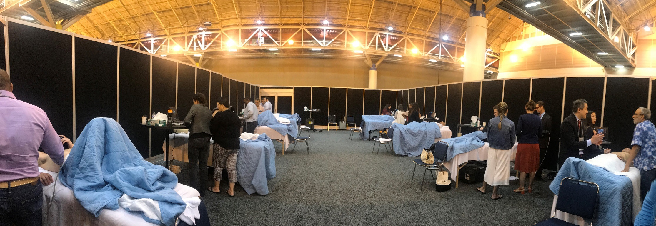
The CHEST Committee has developed a comprehensive panel of hands-on workshops, which are provided by renowned experts in pulmonary, critical care and sleep medicine.
- All Hands-on Workshops are ticketed sessions. Registration for the ticketed sessions will be available during your registration for the congress.
- Each Hands-on Workshop has limited space available and tickets are sold on a first-come first-served basis.
- Since space is limited, please only register for the sessions that you plan to attend. If you have registered for a session that you are unable to attend, kindly let us know straightaway to enable others to participate.
Fundamentals of Bronchoscopy Hands-On Simulation
Description:
This session will review the indications, contraindications, outcomes ad potential adverse events related to fundamental bronchoscopic techniques.
Learning Objectives
- To list the safety issues and techniques of bronchoscopy in special populations: pregnancy, severe asthma, COPD, OSA and heart disease
- To enumerate 4 techniques for managing massive airway bleeding
- To mention the expected results and complications from BAL, brushings, endobronchial and transbronchial biopsy
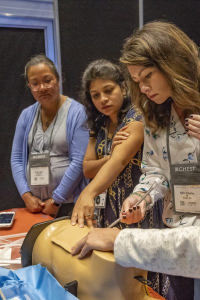
About the workshop
Participants will learn new skills in this hands-on course led by experts in bronchoscopy from Italy and USA. Course content will cover managing massive hemoptysis, foreign body extraction, airway and mediastinal anatomy and cryodebulking.
After participating in this educational activity, the participants should be able to:
- Outline and demonstrate the various techniques for managing massive airway bleeding
- Demonstrate foreign body removal using flexible bronchoscopic techniques: forceps, basket, and cryoprobe
- Define the role and demonstrate the use of endobronchial blockers for massive hemoptysis
- Demonstrate equipment, setup, and techniques for performing cryodebulking bronchoscopy
- Improve cognitive and technical skills pertinent to airway and mediastinal anatomy
Interventional Management of Pleural Diseases Hands-On Simulation
Description
This interactive session is designed to provide and up to date review of best published evidence on interventional management of patients with pneumothorax, infection and malignant pleural disease. This will be accomplished using a case-based review format with an audience response system. Participants will develop a comprehensive knowledge of the indications, contraindications and associated complications of common pleural procedures including ultrasound guided thoracentesis, chest tube and indwelling pleural catheter placement, pleural biopsy and thoracoscopy.
Learning Objectives
- Discuss in detail the evaluation and management of patients with pleural effusion and pneumothorax.
- Develop a comprehensive knowledge of the indications, contraindications and associated complications of common pleural procedures
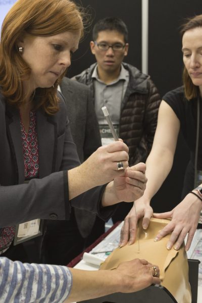
About the workshop
1. Ultrasound guided thoracentesis
a. Learning objectives
- To identify the major anatomical landmarks on ultrasound examination
- To practice thoracentesis using a validated checklist
b. Description
- This station will familiarize learners with the characteristic 2D thoracic ultrasound findings seen in pleural effusion. Learners will become familiar with identifying important anatomic structures such as diaphragm, liver, spleen, and lung margin. The station will include two identical substations, each staffed by one faculty. The model at each station will be a Blue Phantom Thoracentesis Ultrasound Training Model. Once the learners are comfortable with ultrasound identification of key anatomic structures and pleural effusion they will practice simple aspiration of the pleural effusion.
2. Tunneled pleural catheter for effusion
a. Learning Objectives:
- To identify the major anatomical landmarks on ultrasound examination for TPC insertion (identify the triangle of safety)
- To practice TPC insertion step by step by following the dedicated checklist
b. Description:
- This station is designed familiarize learners with the placement of tunneled indwelling pleural catheters. The model at each station is a Simulab box with life like soft tissue. Each model is filled with fluid thus simulating pleural effusions. Learners will identify an appropriate insertion site and will practice placement of tunneled indwelling pleural catheters.
3. Wire guided small bore pleural catheter (pigtail) for pneumothorax
a. Learning Objectives
- Learn the proper steps to insert a small-bore chest tube using percutaneous Seldinger technique
- Practice inserting small bore chest tubes
- Know chest wall landmarks and anatomy for safe insertion of tube
b. Description
- This station is designed to increase the technical knowledge for wire guided small bore chest tube insertion. Learners will become familiar with identifying important anatomic structures such as the 2ndICS in the mid clavicular line. The model at each station will be a Sawbones Thoracostomy model. Using a dedicated check list learners will practice chest tube insertion.
4. Wire guided large bore chest tube
a. Learning Objectives
- Learn the proper steps to insert a large bore chest tube using percutaneous Seldinger technique
- Practice inserting large bore chest tubes
- Know chest wall landmarks and anatomy for safe insertion of tube
b. Description
- This station is designed to increase the technical knowledge for wire guided large bore chest tube insertion. Learners will become familiar with identifying important anatomic structures such as the triangle of safety. The model at each station will be a Sawbones Thoracostomy model. Using a dedicated check list learners will practice chest tube insertion.
Thoracic Ultrasonography Hands-On Scanning
Description
Thoracic ultrasonography is a noninvasive and readily available imaging modality that has important applications in pulmonary medicine. It can be used both diagnostically and also for procedural guidance. A strong application is its use for rapid diagnosis in acute respiratory failure. This session will provide the learner both a basic lecture-based overview of thoracic ultrasonography and then hands-on practice to acquire the ultrasound images with real models.
Learning Objectives
- Understand the knowledge base required to perform and interpret thoracic ultrasonography
- Acquire image acquisition skills required for thoracic ultrasonography
- Understand the use of thoracic ultrasonography for rapid diagnosis of acute respiratory failure
- Understand the difference between A lines, B lines and consolidation pattern
- Understand the characteristics and landmarks of a pleural effusion
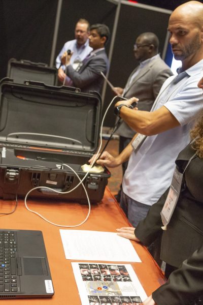
About the workshop
Attendees will experience:
- The scanning technique of examining a thorax in the supine and upright patient
- Knowing the meaning of A lines and how to find them
- Know how to find and optimize lung sliding and know about lung point
- Know B lines and their characteristics
- Know what a consolidation pattern looks like and where to find them
- Know where and how to look for pleural effusion and its basic landmarks
Point of Care Echo Hands-on Simulation
Description
This ultrasonography session will provide a lecture-based overview followed by a hands-on opportunity to practice image acquisition on live models. Specifically, it is designed to focus on the patient with an unknown cause of hemodynamic failure. Image acquisition will be practiced during small-group hands-on workshops. Practice your skills using high-quality ultrasonography technology on live patient models, supervised by experienced faculty who will guide and maximize your training.
Learning Objectives
- Understand the knowledge base required to perform critical care echocardiography and interpret cases
- Incorporate interpretation skills required for critical care ultrasonography and echocardiography
- Demonstrate appropriate image acquisition techniques required for critical care echocardiography
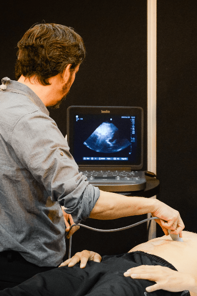
About the workshop
Attendees will experience:
- The optimum position of the patient and how to hold the probe
- Understand basic knobology
- Depth
- Axis for each view
- Gain
- Hand positioning for each of the 5 cardiac views
- Find and set optimum axis and the anatomy of the:
- Parasternal long axis view
- Parasternal short axis view
- Apical 4 chamber view
- Subcostal long axis view
- IVC view
